


In cases of thoracic osteonecrosis, organs involving areas of the spinal cord, located at the level of the affected thoracic region downward, are often affected. Violation of the normal functioning of the spine leads to immobility of the arms, legs and whole body, dysfunction of the pelvic organs, respiratory muscles and internal organs.
Osteoma is a degenerative - dystrophic disease of the spine, the disease is based on changes in the intervertebral discs that are associated with the pathological process of the adjacent vertebrae and disc joints with the entire ligamentous apparatus. .
Features of spinal anatomy
Mobility and stability, elasticity and elasticity of the spine depend largely on intervertebral discs, which are one of the types of cartilaginous connections between bones and create a tight connection between the bodies of the vertebrae. vicinity. The total length of the intervertebral discs is one-quarter of the length of the spine.
The most important function of the disc is to reduce the vertical load on the vertebrae. The disc consists of three parts:
- hyaline plate (closely adjacent to the vertebrae);
- pulp (fills the space between the plates);
- yarn ring (encircling the nucleus from the outside in).
The nucleus contains chondrocytes, tightly interwoven collagen and chondrin (proteoglycan) fibers. The anterior surface of the disc is covered by the anterior longitudinal ligament, which fuses tightly with the vertebrae and flips freely over the disc. The posterior longitudinal ligament is closely fused with the surface of the intervertebral disc and forms the anterior wall of the spinal canal. The disc has no blood supply of its own, so it feeds on substances that come from diffusion from the vertebral bodies.
The vertical distribution of loads in the spine occurs due to the elastic nature of the discs. As a result of the pressure, the pulp nucleus expands, and the pressure is redistributed to the annular and hyaline plates. During locomotion, the core moves in the opposite direction: when bending - to the convex side, when not bending - forward. As the spine moves, the muscles, ligaments, and discs are included in the work. Thus, violations in one link lead to violations in the entire kinetic chain.
Causes and mechanism of development of the disease
During the development of osteonecrosis, mechanical action on the spine plays a special role. Under the influence of unfavorable static and dynamic loads, the core gradually loses its elastic properties (as a result of the polymerization of polysaccharides), forming protrusions and sequestration.
The degenerative disc process is influenced by genetic predisposition, which causes the development of changes in the neuromuscular apparatus of the back, changes in the structure of glycosamines, and a violation of the distribution of collagen fibers in the spine. disc. Genetic factors are paramount in the occurrence of thoracic osteonecrosis, which may increase functional activity.
Risk factors for the development of degenerative changes in the spine include anatomical features of the discs, which are evolutionary imperfections. One of these characteristics is the nutritional character of the structure. In the human body, intervertebral discs are composed of poorly perfused tissues. The closure of blood vessels has occurred during childhood. After nutrition occurs due to the diffusion of substances through the terminal plates.
The factor that stimulates the penetration of nutrients is the load that excludes static postures and great stress. Physical inactivity is one of the leading risk factors for thoracic osteonecrosis. Therefore, regular exercise is an important preventive measure.
The specificity of the microstructure - a few cells - reduces the intensity of the regenerative capacity and the speed of recovery of the disc components. An anatomical feature is the weakness and lack of strength of the posterior intervertebral discs. This contributes to the appearance of wedge-shaped discs in the lower thoracic and lumbar regions.
Great importance in the development of osteonecrosis is given to insignificant changes. Active degenerative changes begin to increase after 30 years. The synthesis of the necessary components for the plate (glycosaminoglycans) continues, but their quality deteriorates. The hydrophilicity decreases, the fiber content increases, the appearance of sclerosis.
Degenerative stages of discs:
- asymptomatic prolongation, degenerative changes in the inner disc components, displacement of the nucleus within the disc;
- pronounced lens symptoms of thoracic osteonecrosis, spinal cord compression, medullary nucleus protrusion (eye protrusion, 1 degree);
- disc rupture with cranial convexity (herniation, grade 2);
- Degenerative changes in the extravertebral disc components (layer 3).
Condylar pathology compresses nerve roots, blood vessels or spinal cord at different levels (cervical, thoracic, lumbar spine) determining the clinical picture.
The limitation of mobility of the thoracic spine, due to the presence of a rib cage, contributes to the least amount of disc trauma, and thus osteonecrosis. Physiological thoracic kyphosis contributes to redistribution of upper body weight to the lateral and anterior portions of the vertebrae. As a result, herniated discs and osteomas form on the front and sides of the spine. Osteosarcoma and posterior hernia are extremely rare.
Osteomas contribute to narrowing of the disc lumen and compression of the roots of the spinal cord and sympathetic fibers. Sympathetic fibers originate in the gray matter of the spinal cord, then aggregate into nodes, from where they are sent to all internal organs. This leads to the fact that osteonecrosis of the thoracic cavity, in addition to typical neurological disorders, also leads to dysfunction of internal organs (vegetative, vasomotor, nutritional) and mimics other diseases. soma. The degenerative features of the thoracic disc explain the difficulties in making the correct diagnosis and treatment.
Symptoms of thoracic osteonecrosis
Osteonecrosis of the chest is more typical in people who lead a sedentary lifestyle. At the same time, there is no stimulating effect of the load on the spine, which contributes to the disruption of disc restoration. The disease develops in people who work for a long time on the computer, stooped, etc. v. such people need to independently perform therapeutic exercises.
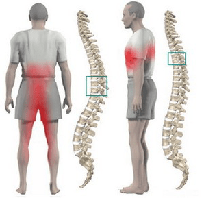
Usually, osteonecrosis of the chest is manifested by dull, less painful and burning pain. Localized pain between the shoulder blades. The patient is disturbed by the feeling of chest compression. When feeling the torsion processes of the thoracic vertebrae, local pain is detected, this pain increases with longitudinal loads on the spine, deep inspiration and body movements.
Some patients experience sharp pain in the collarbone and lower chest (posterior limb syndrome). This symptom develops due to displacement of the lower ribs. Pain increases sharply when rotating the body. More often, the pain syndrome disappears suddenly.
Often, pain is felt in the chest, which corresponds to the movement of the intercostal nerve. Sensitivity in the medial nerve endings is correspondingly disturbed, paresthesia occurs, and often decreases in superficial and deep sensitivities. Possible violation of the function of abdominal presses, changes in knee and muscle tendon reflexes.
Violation of the function of internal organs occurs when any nerve root is compressed at the level of 1 to 12 thorax. In the thoracic region there are structures responsible for nourishing the lungs, heart, intestines, liver, pancreas, and kidneys. Therefore, there are no signs specific to thoracic osteonecrosis.
The disease is manifested by symptoms characteristic of another pathology:
- shortness of breath;
- severe pain at night;
- "heart", angina;
- soreness in the mammary glands;
- pain in the right or left hypogastrium (symptoms of cholecystitis and pancreatitis);
- pain in the throat and esophagus;
- epigastric, abdominal pain (symptoms of gastritis, enteritis and colitis);
- sexual dysfunction.
Diagnose
The greatest value in diagnosing a thoracic osteosarcoma is the chest X-ray examination. The image shows a decrease in disc height, hardening of the end discs, formation of osteoblasts.
Computed tomography allows you to clarify the condition of the vertebrae, joints of the spine, the size of the spinal canal, determine the location of the condyle of the skull and its size.
When performing the differential diagnosis, a careful medical history should be obtained and all clinical signs of thoracic osteonecrosis should be compared with those of other diseases. Examples: nitroglycerin-associated cardiac pain, epigastric pain unrelated to eating, non-seasonal, all symptoms appearing mainly in the evening and completely disappearing after one night's rest.
How to treat thoracic osteonecrosis?
Treatment of osteonecrosis of the thoracic spine in most cases is conservative. Indications for treatment are visceral syndromes with predominant neurological disorders. The primary chiropractic treatment should be adequate traction for the spine:
- underwater active longitudinal traction;
- Passive traction on slant bed using Glisson loop in case of degree 1-4 thoracic vertebrae injury, with axillary strap in case of grade 4-12 thoracic vertebrae injury.
Drug treatment consists of performing a vertebral blockade with a solution of novocaine. With an exacerbation of the disease, analgesics and sedatives are used. With pain syndrome without manifestation, it is possible to use ointments with analgesics and anti-inflammatory drugs at home.
After eliminating acute phenomena, massage of the muscles of the back and lower extremities is used. Manual therapy is indicated for grade 1-3 osteonecrosis in case of development of functional blockade. It includes different options for soft and rough effects on the back muscles.
Therapeutic exercises allow you to load all parts of the spine in doses, which helps to stimulate the recovery process. An important condition for exercise therapy for osteonecrosis is the exclusion of vertical loads.
Physiotherapy: UHF treatment, ultrasound, inductance, radon and pine needle salt soak. At the spa stage, underwater traction and hydromassage are actively used.
Surgical treatment is rarely used. Indication for surgical intervention is spinal cord compression by prolapsed disc fragment.